15.3 Viscodissection in Proliferative Diabetic Retinopathy
Viscodissection of preretinal membranes is useful in the management of tractional retinal detachment (TRD) of several etiologies, notably proliferative diabetic retinopathy (PDR) and sickle cell retinopathy.
In TRD due to PDR, the ideal use for viscodissection is for the removal of diffuse preretinal membranes with tight and/or numerous retinal adhesions. Viscodissection serves to create a surgical plane in the potential space between the preretinal membranes and the retina, “holding” the retina posteriorly while separating and “lifting” the preretinal membranes. Further advantages include the loculation of preretinal hemorrhaging by the viscoelastic and the ability to stabilize retina that is both tractionally and rhegmatogenously detached.
The Hubbard Viscodissector (Bausch & Lomb) was created to facilitate viscodissection of posterior and peripheral retinal membranes. It is available in both 25- and 27-gauge. It features an extendable cannula that angles to 90 degrees. It is used in conjunction with the viscous fluid injection syringe and tubing and the viscous injection settings on the vitrectomy machine.
Figure 15.3.1 and Figure 15.3.2 The Hubbard Viscodissector for Viscodissection of Preretinal Membranes
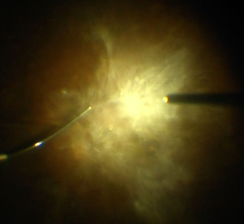
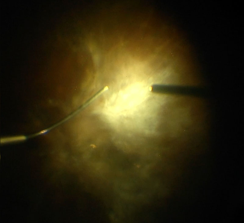
Figure 15.3.1 and Figure 15.3.2 The Hubbard Viscodissector for Viscodissection of Preretinal Membranes
Setup and Procedure
- Obtain two tubes of the viscoelastic of choice (a cohesive viscoelastic is preferred) and load them into a 10cc/ml syringe, with the VFC plunger closed to approximately 1.5cc/ml. Viscoelastic is carefully loaded to ensure no air is present. The proper technique involves injecting the viscoelastic to the side of the syringe while twirling the syringe in your hand almost in the same fashion as a soft serve ice cream
- The viscodissection is then attached first to a short length flexible tubing (short to prevent excessive viscoelastic waste) and then the viscodissector
- The viscodissector is then primed at an appropriate injection pressure (typically 30-38 mmHg)
- Before introducing the viscodissector into the eye, a limited core vitrectomy is performed to enhance visualization and remove vitreous hemorrhage. The anterior and posterior hyaloid should be separated
- A posterior hyaloidotomy must be identified or created so that the viscodissector can be inserted between the posterior hyaloid and the retina. Ideally, this hyaloidotomy is left small to prevent excessive backflush of injected viscoelastic. The vitrector is then removed and the viscodissector is inserted. The cannula is placed between the posterior hyaloid (and intrinsic membranes) and retina and the tip extended until it is parallel with the retinal surface. This facilitates separation of preretinal membranes and decreases the chance of retinal break and inadvertent subretinal viscoelastic injection (Figure 15.3.1)
- Viscoelastic is then injected using the automated, foot-pedal control. The viscoelastic then dissects between the preretinal membranes and the retina, creating the surgical plane (Figure 15.3.2)
- The tip of the viscodissector can be moved laterally to perform blunt dissection of identified tight vitreoretinal fibrovascular adherent pegs
- If a significant but subtotal amount of preretinal membranes is dissected, the viscodissector can be exchanged for the vitrector and separated preretinal membranes can be safely removed without involving the retina (Figure 15.3.3). The viscodissector is then reinserted to address further preretinal membranes
All rights reserved. No part of this publication which includes all images and diagrams may be reproduced, distributed, or transmitted in any form or by any means, including photocopying, recording, or other electronic or mechanical methods, without the prior written permission of the authors, except in the case of brief quotations embodied in critical reviews and certain other noncommercial uses permitted by copyright law.

Westmead Eye Manual
This invaluable open-source textbook for eye care professionals summarises the steps ophthalmologists need to perform when examining a patient.