13 Rhegmatogenous Retinal Detachment Special Scenarios
13.1 Rhegmatogenous Retinal Detachment: Re-detachment Surgery
13.2 Macular Hole Retinal Detachment
13.3 Retinoschisis Retinal Detachment
13.4 Optic Disc Pit Retinal Detachment and Maculopathy
13.5 Giant Retinal Tear Detachment
13.6 Retinal Dialysis
13.7 Macular Folds
13.8 Sickle Cell Detachment
13.9 Viral Retinitis Associated Retinal Detachment
13.10 Paediatric Retinal Detachment
13.11 Coloboma Associated Retinal Detachment
13.12 Inherited Retinal Dystrophies and Retinal Detachment
13.7 Macular Folds
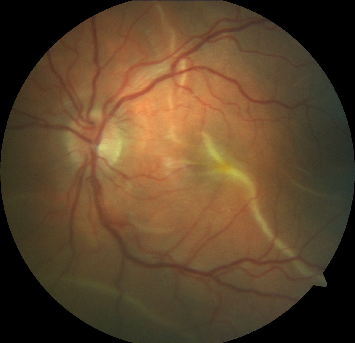
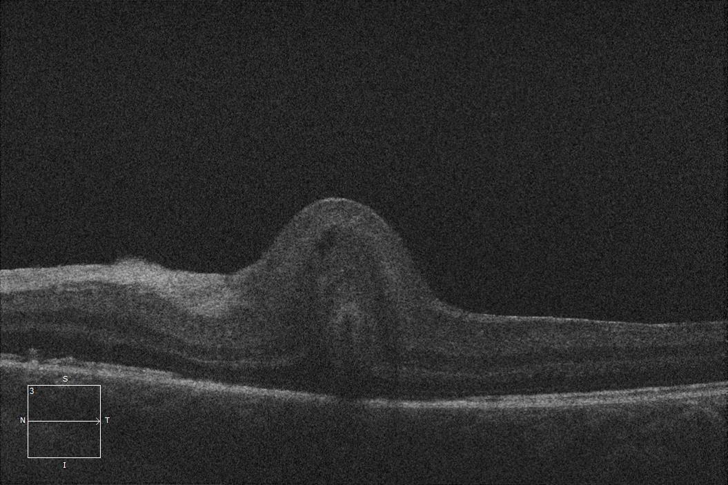
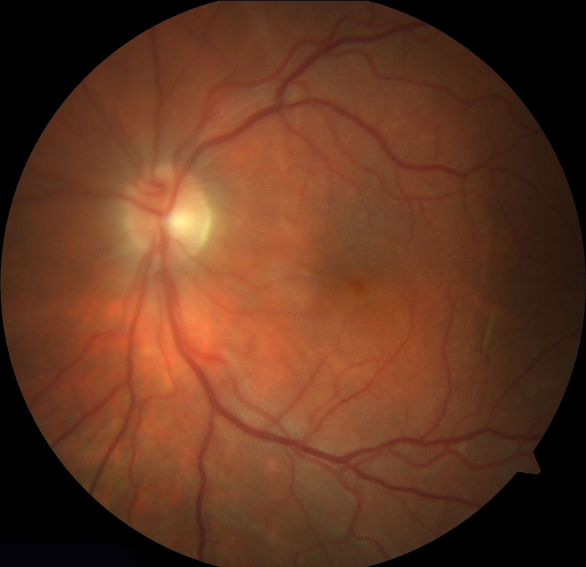
Macular retinal folds can occur following any retinal detachment surgery and are considered a result of residual sub-retinal fluid (SRF) and incomplete positioning (Figure 13.7.1 and Figure 13.7.2). Folds are probably caused by accumulation of SRF in a gravity-dependent position. Risk factors include superior bullous detachment, macula off detachment where drainage has occurred from the break and residual SRF remains, circumferential buckles and from slippage in giant retinal tears.[1,2,3] Non-foveal folds probably have little clinical significance. When macular retinal folds are observed a re-operation should be scheduled as soon as practicable to unfold the retina as outer retinal denegation occurs as early as 1 week after detachment. Foveal folds are usually associated with poor vision and do not improve spontaneously, but the visual improvement can be significant following successful unfolding. While surgery can sometimes be successful in resolving a macular fold, the best practice is to avoid them by performing complete drainage in cases at higher risk of fold (with either perfluoro-n-octane or a posterior drainage retinotomy) and in rapidly positioning the patient in the face down position as soon as surgery has been completed (Figure 13.7.3 and Figure 13.7.4).
Heimann H, Bopp S. Retinal folds following retinal detachment surgery. Ophthalmologica 2011;226 Suppl 1:18-26.
Witkin AJ, Hsu J. Surgical repair of macular fold after vitrectomy for bullous rhegmatogenous retinal detachment. Retina 2012;32(8):1666-9.
Zacharias LC, Nóbrega PF, Takahashi WY. Surgical correction of retinal folds involving the fovea. Ophthalmic Surg Lasers Imaging Retina 2014;45(1):50-3.
Previous
13.6 Retinal Dialysis
All rights reserved. No part of this publication which includes all images and diagrams may be reproduced, distributed, or transmitted in any form or by any means, including photocopying, recording, or other electronic or mechanical methods, without the prior written permission of the authors, except in the case of brief quotations embodied in critical reviews and certain other noncommercial uses permitted by copyright law.

Westmead Eye Manual
This invaluable open-source textbook for eye care professionals summarises the steps ophthalmologists need to perform when examining a patient.