5.1 Intraocular Tamponades and Vitreous Substitutes
Multiple different intraocular tamponades exist. Their properties including buoyancy, expansibility, maximum size, duration, viscosity, toxicity and arc of contact are outlined in Tables 1 and 2. These properties will determine the best choice of tamponade in different clinical scenarios (Table 1) [1,2,3,4,5]
Singh RP. Global Trends in Retina Survey. Chicago, Illinois: American Society of Retina Specialists; 2018 [cited 2019 September 25].
Foster WJ, Chou T. Physical mechanisms of gas and perfluoron retinopexy and sub-retinal fluid displacement. Phys Med Biol. 2004;49(13):2989-97.
Schachat AP, Wilkinson CP, Hinton DR, Wiedemann P, Freund KB, Sarraf D, et al. Ryan's Retina; 2017.
Chan CK, Lin SG, Nuthi AS, Salib DM. Pneumatic retinopexy for the repair of retinal detachments: a comprehensive review (1986-2007). Surv Ophthalmol. 2008;53(5):443-78.
Mohamed S. Intraocular gas in vitreoretinal surgery. Hong Kong Journal of Ophthalmology. 2010;14:8-13.
Physical Properties
Duration
Largest Size
Expansion (Pure)
Indications
Air
Physical Properties
- Floats
Duration
5 - 7 days
Largest Size
Immediate
Expansion (Pure)
No expansion
Indications
- Tamponade of sclerostomies for routine cases (e.g. epiretinal membrane surgery)
SF6
Physical Properties
- Floats
- 20% Non-expansile
- 26% if some expansion desired
Duration
1 - 2 weeks
Largest Size
36 hours
Expansion (Pure)
2X
Indications
- Non-PVR, primary RRD
- Routine macular hole surgery
C3F8
Physical Properties
- Floats
- 12 - 14% Non-expansile
- 16 - 18% if some expansion desired
Duration
6 - 8 weeks
Largest Size
3 days
Expansion (Pure)
4X
Indications
- Inferior breaks (compared with SF6)
- Mild to moderate PVR retinal detachment
- Giant retinal tear
- Some chronic macular hole surgery
Silicone Oil
Physical Properties
- Floats
- Non-expansile
- 1000 - 10000 centistoke
Duration
Until removed
Largest Size
Immediate
Expansion (Pure)
No expansion
Indications
- Moderate to severe PVR retinal detachment
- Giant retinal tear
- Air travel
- Re-operations
Heavy Silicone Oil
Physical Properties
- Sinks
- Non-expansile
- Viscosity 1400mPas
- Retinotoxic after 3 months
Duration
Until removed (best <3 months)
Largest Size
Immediate
Expansion (Pure)
No expansion
Indications
- Moderate to severe PVR retinal detachment (especially with inferior pathology)
- Giant retinal tear
- Air travel
- Re-operations
Perfluoro-n-octane (PFO)
Physical Properties
- Sinks
- Non-expansile Retinotoxic after 2 weeks
Duration
Until removed (best <2 weeks)
Largest Size
Immediate
Expansion (Pure)
No expansion
Indications
- Intra-operative “third hand” to reattach the retina or float a foreign body or dropped nucleus.
- Temporary endotamponade of severe PVR or a giant retinal tear
Table 1. Air/Gas Properties
Arc of Contact (º)
Gas Bubble Volume (mL/cc)
90
120
150
180
0.28
0.75
1.49
2.40
Table 2. Arc of Contact for Gas Tamponades
Buoyancy and surface tension[2] are the characteristics of intraocular gas which help retinal reattachment after surgery. The buoyant force pushes the retina upward whereas the surface tension prevents flow of fluid into the subretinal space. Choosing appropriate air/gas is depended on indications summarized in Table 1 and 2.[3,4] The decision on tamponade should be made based on an assessment of the location and type of pathology, the expected intraoperative fill and the ability of the patient to position. Intraoperative fill is affected by the degree of vitreous removal, the intraocular pressure at closure and any sclerostomy leakage. Though the approximate duration of each tamponade is listed in the table below, results can vary based on multiple factors.
An intraocular gas bubble has the following dynamics: expansion, equilibration, and dissolution.[5] Expansion is due to diffusion of nitrogen to reach equilibrium. Therefore, it is very important to turn off a nitrogen infusion during general anaesthesia, if used, while performing vitrectomy with gas tamponade.
Foster WJ, Chou T. Physical mechanisms of gas and perfluoron retinopexy and sub-retinal fluid displacement. Phys Med Biol. 2004;49(13):2989-97.
Schachat AP, Wilkinson CP, Hinton DR, Wiedemann P, Freund KB, Sarraf D, et al. Ryan's Retina; 2017.
Chan CK, Lin SG, Nuthi AS, Salib DM. Pneumatic retinopexy for the repair of retinal detachments: a comprehensive review (1986-2007). Surv Ophthalmol. 2008;53(5):443-78.
Mohamed S. Intraocular gas in vitreoretinal surgery. Hong Kong Journal of Ophthalmology. 2010;14:8-13.
The gas should be drawn with a sterile filter to prevent microbial contamination. Repetitive air flush out from the syringe can be performed to decrease residual air which might dilute the obtained gas concentration. There are two choices for intravitreal gas injection- a pure (100%, expansile) or percentage (non-expansile) draw:
Pure Draw
This is mainly used for pneumatic retinopexy, when a vitrectomy has not been performed. A small amount of gas (e.g. 0.3 - 0.5 ml of 100% SF6 or 100% C3F8) is injected intravitreally. This will expand over the following days.
Percentage Draw
With a percentage draw, the actual percentage of gas desired is injected in the eye. A necessary volume of 100%-filtered gas is drawn into a 50 - ml syringe. Filtered air is then drawn into the same syringe until the volume in the syringe reaches 50 ml. For instance, for a 20% SF6 mixture, 10 ml of 100% SF6 is drawn up to 50 ml by adding 40 ml of air. For a 14% C3F8 mixture, 7 ml of 100% C3F3 is drawn up to 50 ml by adding 43 ml of air. Approximately 40 ml of the gas mixture is injected into the intravitreal cavity. Some gas products come in pre-mixed, non-expansile concentrations (e.g. EasyGas®).
Note
- Gas should not be drawn up too far in advance to avoid dilution errors. If drawn up a few minutes in advance, it should be stored with the end of the syringe pointing up since gas is heavier than air
- Always place the filter closest to the syringe that the gas is being drawn up into
There are multiple methods for injecting intravitreal gas: Ensure complete drainage of fluid from the eye (e.g after retinal detachment or macular hole surgery) to ensure a maximum gas fill.
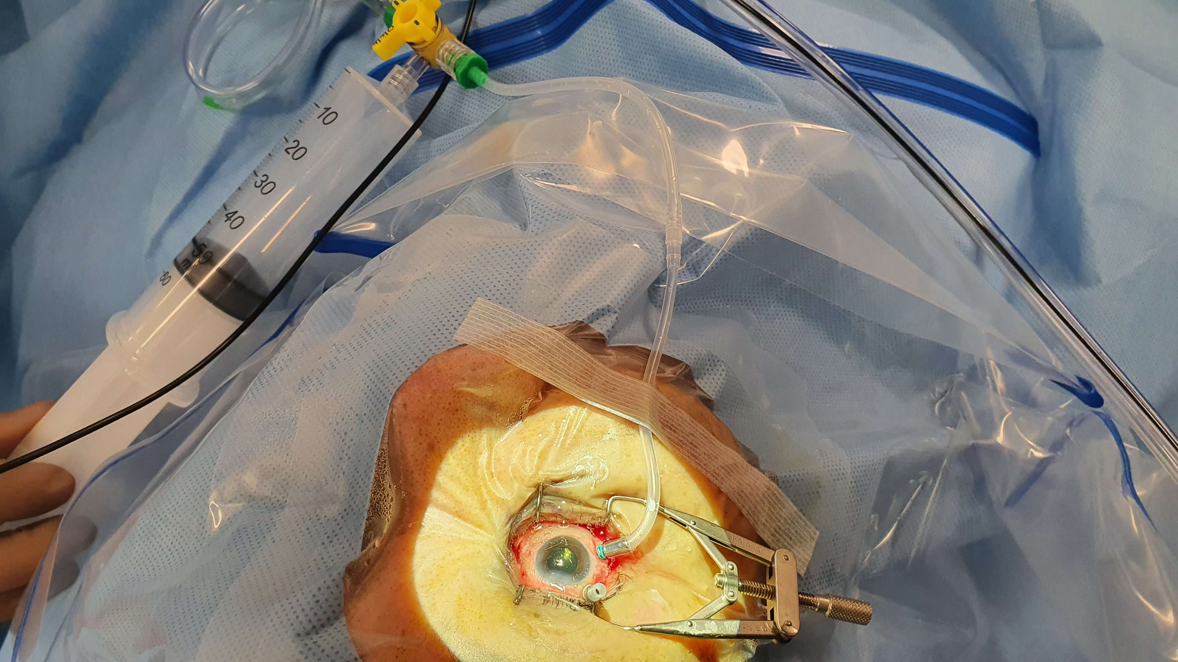
Method 1- Injecting from the Infusion Line
(Figure 5.1.1)
- Remove one of the superior cannulas. Ensure that this is not leaking- suture it if it is
- Ensure that the remaining superior cannula is open. This will be the case if the cannula is non-valved. If it is valved, it will need to be opened. Options include inserting the vent that comes with the vitrectomy pack into the cannula, inserting a chimney or placing forceps into the cannula to open the valve. If both superior cannulae have been removed, a needle inserted through the pars plana can be used to vent gas
- Attach the syringe of gas to the 3-way stopcock on the infusion line and turn the stopcock so that it is open towards the eye but closed towards the vitrectomy machine. If there is no 3-way stopcock, the syringe of gas will need to be connected directly to the infusion line (clamp the infusion whilst it is disconnected and reconnected to the syringe of gas to avoid hypotony). During this process, be careful as the eye may deflate as there is no longer any infusion pressure
- Slowly inject the gas from the infusion line, venting it out the remaining superior sclerostomy. This is usually performed by an assistant who will call out every 5ml/cc reduction of gas in the syringe. The gas mixture will displace out the air in the eye. Stop injecting when there is approximately 10ml/cc of gas remaining in the syringe. At this point the surgeon will remove the superior sclerostomy cannula. The IOP can be checked digitally, and if too low a small amount of supplemental gas can be injected (this is the reason for reserving 10ml/cc of gas in the syringe). The infusion line is removed last
Method 2- Injecting from the Sclerostomy Cannula
- Remove one of the superior cannulas. Ensure that this is not leaking- suture it if it is
- Open the infusion line to atmospheric, either by disconnecting it from the vitrectomy machine or turning the 3-way stopcock so that it is open to atmospheric
- Slowly inject the gas from the remaining superior sclerostomy, venting it out the infusion line. Condensation within the infusion line may be visualized as the warmer air from the eye is displaced out. Remove the superior sclerostomy cannula then the infusion line cannula
Method 3- Injecting from the Pars Plana
- Remove both of the superior cannulae. Ensure that they are not leaking- suture them if they are
- Slowly inject the gas intravitreally using a 30-gauge needle on the 50ml/cc syringe, venting it out the infusion line. The needle is inserted into the intravitreal cavity at an IOP of 20 - 25 mmHg so that the globe has some resistance, before the infusion line 3-way stopcock is opened to atmospheric pressure or the infusion line is disconnected. Condensation within the infusion line may be visualized as the warmer air from the eye is displaced out. After injecting the gas, remove the infusion line cannula
Note
- If the gas is being injected through a 20-gauge vitrectomy, there is a risk of hypotony and loss of gas prior to closure. If this is required, the sclerotomy will need to be sutured closed. This is best pre-placed to minimize the time for closure
- All patients who have intravitreal gas should wear a gas wrist-band post-operatively until the gas has completely dissolved. This serves as a warning to anaesthesiologists/anaesthetists in the event that general anaesthesia is required (nitrous oxide anaesthesia is contraindicated with intravitreal gas)
Silicone Oil Injection
Indications for using SO are listed in the Table 3. The principle steps for infusion and removal are discussed below (Figure 5.1.2).
- If the eye is aphakic, perform an inferior peripheral iridectomy with the cutter to prevent pupil block
- Remove one of the superior cannulae (usually on the side of the non-dominant hand) and suture the sclerostomy closed
- Inject the oil directly through the remaining superior cannula using the viscous fluid control (VFC) syringe. There are attachments that fit either into or over the cannula. Those that fit into the cannula are easier to control but slower to inject due to their smaller internal diameter. Those that fit over the cannula are faster to inject but often slip off the cannula. The larger the cannula, the faster the oil can be injected
- If there is concern that the cannula could be accidentally positioned in the suprachoroidal or subretinal space, the oil injection should be directly visualized
- The infusion line will need to be disconnected, or the 3-way stopcock turned so that it is open to atmospheric pressure. This prevents the IOP from rising as the silicone oil is injected, but it may cause temporary hypotony until the eye is sufficiently filled with silicone oil. An assistant can adjust the IOP by opening and closing the infusion line to atmospheric as the silicone oil is injected. An alternative method is to keep the opposite superior sclerostomy in situ whilst the silicone oil is injected to vent air. This is then removed and the sclerostomy sutured once the eye has sufficient oil fill that it won’t collapse when the infusion line is opened to atmospheric pressure
- As one approaches a complete oil fill, the eye should be tilted so that the cannula that is venting air (usually the infusion line that is open to atmospheric pressure) is positioned most superior. This means that air will be able to escape from the eye and a complete oil fill will be achieved. Continue the infusion until the oil just begins to come up the infusion line and there is no air left in the vitreous cavity. In an aphakic eye, watch for the oil fill to reach the pupil plane but do not over-fill otherwise oil will enter the anterior chamber. It is sometimes useful to have some air in the anterior chamber to prevent oil entering
- Check the IOP by digitally palpating the globe or use a Schiötz tonometer. It is important not to over-fill the eye as it may cause ocular hypertension which is difficult to manage without surgical removal of oil. Less silicone oil is required for superior compared with inferior pathology
- Remove all remaining cannulae and suture all sclerostomies with 7-0 vicryl to prevent subconjunctival oil
All rights reserved. No part of this publication which includes all images and diagrams may be reproduced, distributed, or transmitted in any form or by any means, including photocopying, recording, or other electronic or mechanical methods, without the prior written permission of the authors, except in the case of brief quotations embodied in critical reviews and certain other noncommercial uses permitted by copyright law.
Westmead Eye Manual
This invaluable open-source textbook for eye care professionals summarises the steps ophthalmologists need to perform when examining a patient.


