13 Rhegmatogenous Retinal Detachment Special Scenarios
13.1 Rhegmatogenous Retinal Detachment: Re-detachment Surgery
13.2 Macular Hole Retinal Detachment
13.3 Retinoschisis Retinal Detachment
13.4 Optic Disc Pit Retinal Detachment and Maculopathy
13.5 Giant Retinal Tear Detachment
13.6 Retinal Dialysis
13.7 Macular Folds
13.8 Sickle Cell Detachment
13.9 Viral Retinitis Associated Retinal Detachment
13.10 Paediatric Retinal Detachment
13.11 Coloboma Associated Retinal Detachment
13.12 Inherited Retinal Dystrophies and Retinal Detachment
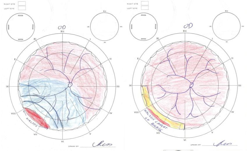
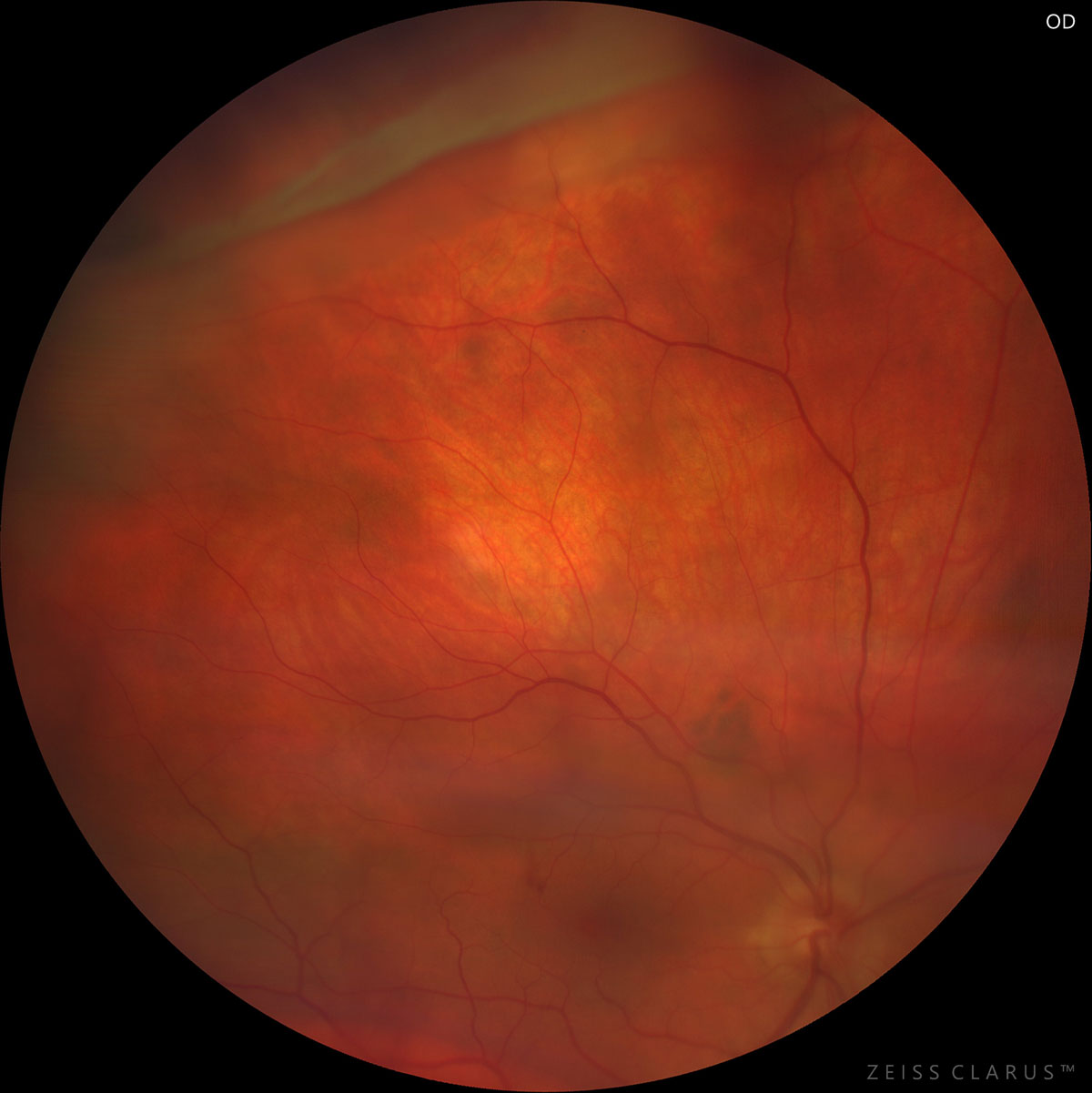
13.6 Retinal Dialysis
Retinal dialysis refers to detachment of the retina from the ora serrata. Over two-thirds occur secondary to blunt ocular trauma[1] and are therefore most common in young males. A retinal detachment may be present as well. The mechanism of these detachments differs from rhegmatogenous retinal detachment in the sense that the vitreous is typically attached in these eyes and the vitreous base is attached to the posterior portion of the retinal break. This gives rise to other typical characteristics of these cases: these detachments progress slowly and often present with demarcation lines[1] and the diagnosis is delayed over one month in 80% of cases. Three-quarters occur in the inferotemporal quadrant. Retinal dialysis should be treated with photocoagulation or cryopexy as prophylaxis for retinal detachment. Scleral buckling with a tire (e.g. no. 286 or 287) remains the treatment of choice for retinal detachment secondary to retinal dialysis, with up to a 98% reattachment rate. External drainage of subretinal fluid is usually not required unless the detachment is very extensive. Cryotherapy should be applied to the anterior horns and the posterior edge of the dialysis. The element should be placed to support the break, which will be relatively anterior at the vitreous base.
Chang JS, Marra K, Flynn HW, Jr., et al. Scleral Buckling in the Treatment of Retinal Detachment Due to Retinal Dialysis. Ophthalmic Surg Lasers Imaging Retina 2016;47(4):336-40.
Next
13.7 Macular Folds
All rights reserved. No part of this publication which includes all images and diagrams may be reproduced, distributed, or transmitted in any form or by any means, including photocopying, recording, or other electronic or mechanical methods, without the prior written permission of the authors, except in the case of brief quotations embodied in critical reviews and certain other noncommercial uses permitted by copyright law.

Westmead Eye Manual
This invaluable open-source textbook for eye care professionals summarises the steps ophthalmologists need to perform when examining a patient.