16 Vitreomacular Interface Disorders
16.1 Epiretinal Membrane Peel
16.2.1 Macular Hole Repair (Surgical)
16.2.2 Macular Hole Repair (Medical Therapy)
16.3 Vitreomacular Traction Syndrome
16.4 Myopic Traction Maculopathy – Vitrectomy
16.5.1 Myopic Traction Maculopathy – Macular Buckling
16.5.2 Myopic Traction Maculopathy – Macular Buckling (AJL)
16.1 Epiretinal Membrane Peel
Epiretinal membrane (ERM) is a disorder of the vitreomacular interface with proliferation of fibrocellular tissue on the surface of the internal limiting membrane (ILM) (Figure 16.1.1). The causes can be idiopathic, secondary to previous vitreoretinal surgery (e.g. retinal detachment surgery), cryotherapy, laser photocoagulation, trauma, uveitis, vitreous hemorrhage, or retinal vascular diseases.[1] Posterior vitreous detachment (PVD) is present in around 95% of idiopathic ERM.[2] Patients can be asymptomatic or present with decreased visual acuity, metamorphopsia, micropsia or macropsia, or distortion of images.
Fung AT, Galvin J, Tran T. Epiretinal Membrane: A Review. Clin Experiment Ophthalmol. 2021;1-20.
Wiznia RA. Posterior vitreous detachment and idiopathic preretinal macular gliosis. Am J Ophthalmol 1986; 102 (2): 196-198.
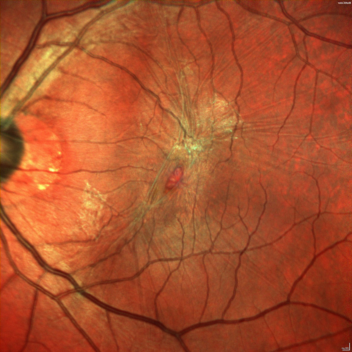
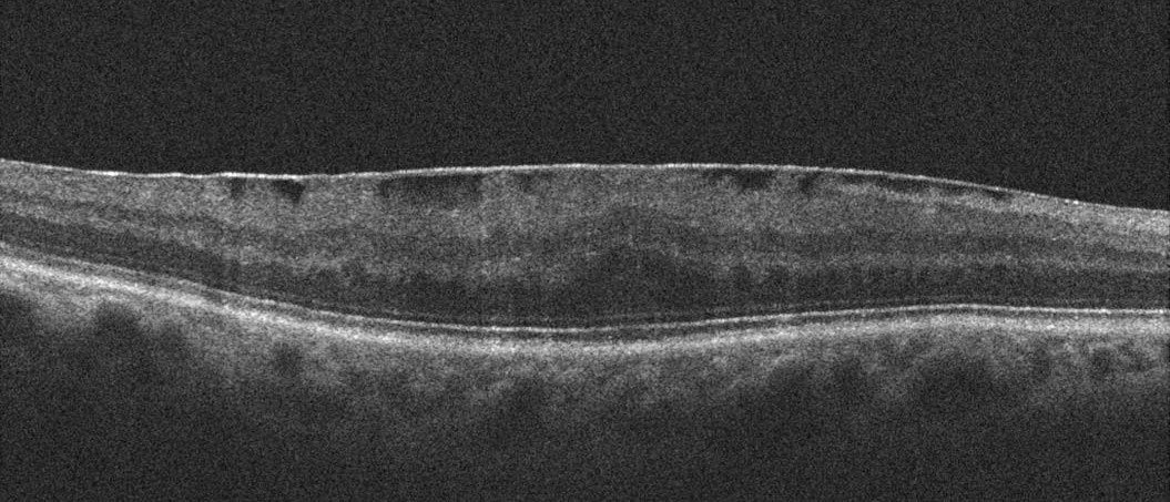
Optical coherence tomography (OCT) is the most sensitive tool for diagnosing ERM (Figure 16.1.2 and Figure 16.1.3).[3] ERM can have focal points of attachment to the retina or generalized diffuse adherence to the retina.[4] There is no universally accepted classification for idiopathic ERM. Based on biomicroscopy, Gass proposed the severity of ERM based on the following grades: Grade 0 (cellophane maculopathy, translucent membranes not associated with any retinal distortion); Grade 1 (crinkled cellophane maculopathy, membranes causing disorganized wrinkling of the inner retina); and Grade 2 (macular pucker, opaque membranes causing distortion of all retinal layers with obscuration of the underlying retinal vessels).[5]
Do DV, Cho M, Nguyen QD, Shah SM, Handa JT, Campochiaro PA, Zimmer-Galler I, Sung JU, Haller JA. The impact of optical coherence tomography on surgical decision making in epiretinal membrane and vitreomacular traction. Trans Am Ophthalmol Soc 2006; 104: 161–166.
Wilkins JR, Puliafito CA, Hee MR, et al. Characterization of epiretinal membranes using optical coherence tomography. Ophthalmology. 1996;103(12):2142–2151.
Gass JDM. Stereoscopic atlas of macular disease. St Louis, Mo: Mosby Year Book; 1987: 716-717.
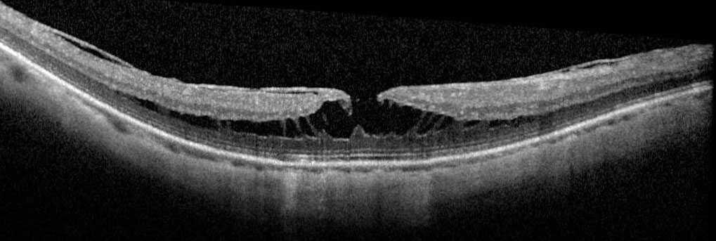
More recently, various OCT-based classification systems for ERM have been proposed based on the foveal morphology, morphology of the macula and vitreoretinal interface, quantitative measurements of central foveal thickness and characterization of inner segment ellipsoid band.[6]
Govetto A, Lalane RA 3rd, Sarraf D, Figueroa MS, Hubschman JP. Insights Into Epiretinal Membranes: Presence of Ectopic Inner Foveal Layers and a New Optical Coherence Tomography Staging Scheme. Am J Ophthalmol. 2017 Mar;175:99-113.
OCT-based ERM Classification by Govetto et al. (Figure 16.1.4) [5]
Figure 16.1.4 Govetto OCT Classification of Epiretinal Membranes
A: Stage 1 (preservation of the foveal dip)
B: Stage 2 (loss of the foveal dip)
C: Stage 3 (ectopic inner foveal layers)
D: Stage 4 (disorganization of the retinal layers)
All rights reserved. No part of this publication which includes all images and diagrams may be reproduced, distributed, or transmitted in any form or by any means, including photocopying, recording, or other electronic or mechanical methods, without the prior written permission of the authors, except in the case of brief quotations embodied in critical reviews and certain other noncommercial uses permitted by copyright law.

Westmead Eye Manual
This invaluable open-source textbook for eye care professionals summarises the steps ophthalmologists need to perform when examining a patient.