2.5 Intraoperative OCT
Over the past decade, optical coherence tomography (OCT) has evolved from a diagnostic imaging modality used exclusively in the clinic to a tool that can be utilized in the operating room.[1] Intraoperative OCT (iOCT) allows the vitreoretinal surgeon to visualize detailed retinal anatomy during surgery beyond the limited en-face view of the retina provided by standard ophthalmic microscopes.[2] The iOCT systems that are currently available create 2D B-scan images in real-time that are useful for visualization immediately before and after surgical manoeuvres.[3] Future developments in swept-source 4D microscope-integrated OCT (MIOCT) may provide a real-time 3D view of retinal structures that facilitate more advanced manoeuvres.[4,5]
The vitreoretinal surgical trainee can effectively utilize the iOCT by following these steps:
Tao YK, Ehlers JP, Toth CA, Izatt JA. Intraoperative spectral domain optical coherence tomography for vitreoretinal surgery. Opt Lett. 2010 Oct 15;35(20):3315-7. doi: 10.1364/OL.35.003315. PMID: 20967051; PMCID: PMC3123722.
Khan M, Ehlers JP. Clinical utility of intraoperative optical coherence tomography. Curr Opin Ophthalmol. 2016 May;27(3):201-9. doi: 10.1097/ICU.0000000000000258. PMID: 26918786; PMCID: PMC4936526.
Ehlers JP. Intraoperative optical coherence tomography: past, present, and future. Eye (Lond). 2016 Feb;30(2):193-201. doi: 10.1038/eye.2015.255. Epub 2015 Dec 18. PMID: 26681147; PMCID: PMC4763135.
Carrasco-Zevallos, O.M., et al., Live volumetric (4D) visualization and guidance of in vivo human ophthalmic surgery with intraoperative optical coherence tomography. Sci Rep, 2016. 6: p. 31689.
Viehland C, Keller B, Carrasco-Zevallos OM, Nankivil D, Shen L, Mangalesh S, Viet du T, Kuo AN, Toth CA, Izatt JA. Enhanced volumetric visualization for real time 4D intraoperative ophthalmic swept-source OCT. Biomed Opt Express. 2016 Apr 12;7(5):1815-29. doi: 10.1364/BOE.7.001815. PMID: 27231623; PMCID: PMC4871083.
Patients requiring procedures with premacular or submacular manipulation may be good candidates for iOCT use.[6] iOCT allows the surgeon to visualize structural changes in the retina after a surgical manoeuvre that may otherwise be invisible with the en-face viewii. For example, the iOCT can confirm that a membrane has been successfully been peeled, an iatrogenic macular hole has not been created, or that subretinal fluid has been adequately drained. It can also confirm that an instrument is in the desired location prior to performing a surgical technique, e.g. accessing the subretinal space for drug delivery (Figure 2.5.1). Vitreoretinal trainees may find iOCT to be useful when performing the following techniques.[7,8,9,10,11]
Pichi F, Alkabes M, Nucci P, Ciardella AP. Intraoperative SD-OCT in macular surgery. Ophthalmic Surg Lasers Imaging. 2012 Nov-Dec;43(6 Suppl):S54-60. doi: 10.3928/15428877-20121001-08. PMID: 23357325.
Ehlers JP, Han J, Petkovsek D, Kaiser PK, Singh RP, Srivastava SK. Membrane Peeling-Induced Retinal Alterations on Intraoperative OCT in Vitreomacular Interface Disorders From the PIONEER Study. Invest Ophthalmol Vis Sci. 2015 Nov;56(12):7324-30. doi: 10.1167/iovs.15-17526. PMID: 26559478; PMCID: PMC4642608.
Ehlers JP, Kernstine K, Farsiu S, Sarin N, Maldonado R, Toth CA. Analysis of pars plana vitrectomy for optic pit-related maculopathy with intraoperative optical coherence tomography: a possible connection with the vitreous cavity. Arch Ophthalmol. 2011 Nov;129(11):1483-6. doi: 10.1001/archophthalmol.2011.316. PMID: 22084218; PMCID: PMC3509483.
Ehlers JP, Petkovsek DS, Yuan A, Singh RP, Srivastava SK. Intrasurgical assessment of subretinal tPA injection for submacular hemorrhage in the PIONEER study utilizing intraoperative OCT. Ophthalmic Surg Lasers Imaging Retina. 2015 Mar;46(3):327-32. doi: 10.3928/23258160-20150323-05. PMID: 25856818; PMCID: PMC4442641.
Gregori NZ, Lam BL, Davis JL. Intraoperative Use of Microscope-Integrated Optical Coherence Tomography for Subretinal Gene Therapy Delivery. Retina. 2019 Oct;39 Suppl 1:S9-S12. doi: 10.1097/IAE.0000000000001646. PMID: 28426632.
Wykoff CC, Berrocal AM, Schefler AC, Uhlhorn SR, Ruggeri M, Hess D. Intraoperative OCT of a full-thickness macular hole before and after internal limiting membrane peeling. Ophthalmic Surg Lasers Imaging. 2010 Jan-Feb;41(1):7-11. doi: 10.3928/15428877-20091230-01. PMID: 20128563.
- Membrane peeling
- Macular hole closure (Figure 2.5.2)
- Vitreomacular traction release
- Traction retinal detachment repair
- Subretinal drug delivery
- Examination under anaesthesia
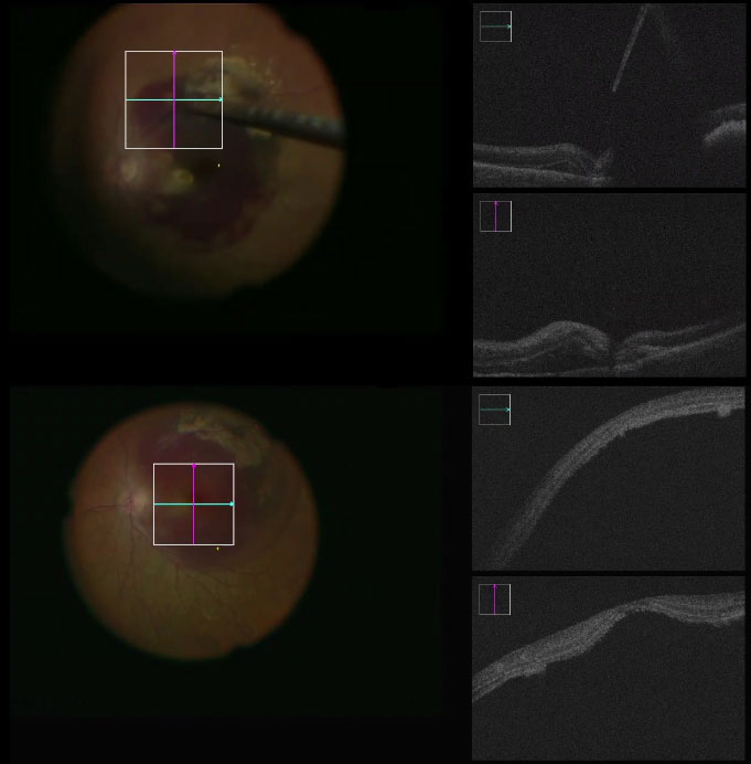
Figure 2.5.1 Submacular tPA Injection using Zeiss RESCAN iOCT
Top: En-face view (left). 0° and 90° iOCT B-scans (right). The 41-gauge needle (arrow) penetrates into the subretinal space. Note that the instrument causes significant shadowing artifact in the 2D image (star)
Bottom: iOCT demonstrating successful delivery of tPA into the subretinal space
i. Hand-held OCT (Figure 2.5.3)
A hand-held 2D OCT system is now commercially available as the Envisu C-Class (Leica Microsystems).[12] The device is particularly useful in non-sterile cases such as examinations under anaesthesia in young children or other patients who cannot undergo OCT imaging in a clinic setting.[8] It can also be used during vitreoretinal surgery under sterile conditions; however, it requires a pause in the surgery, removal of the microscope, and placement of a sterile bag over the imaging handpiece.[3] Despite these limitations, hand-held OCT can be useful when microscope-integrated OCT is not readily available.
Dayani PN, Maldonado R, Farsiu S, Toth CA. Intraoperative use of handheld spectral domain optical coherence tomography imaging in macular surgery. Retina. 2009 Nov-Dec;29(10):1457-68. doi: 10.1097/IAE.0b013e3181b266bc. Erratum in: Retina. 2011 Jun;31(6):1236. PMID: 19823107; PMCID: PMC3515871.
Ehlers JP, Kernstine K, Farsiu S, Sarin N, Maldonado R, Toth CA. Analysis of pars plana vitrectomy for optic pit-related maculopathy with intraoperative optical coherence tomography: a possible connection with the vitreous cavity. Arch Ophthalmol. 2011 Nov;129(11):1483-6. doi: 10.1001/archophthalmol.2011.316. PMID: 22084218; PMCID: PMC3509483.
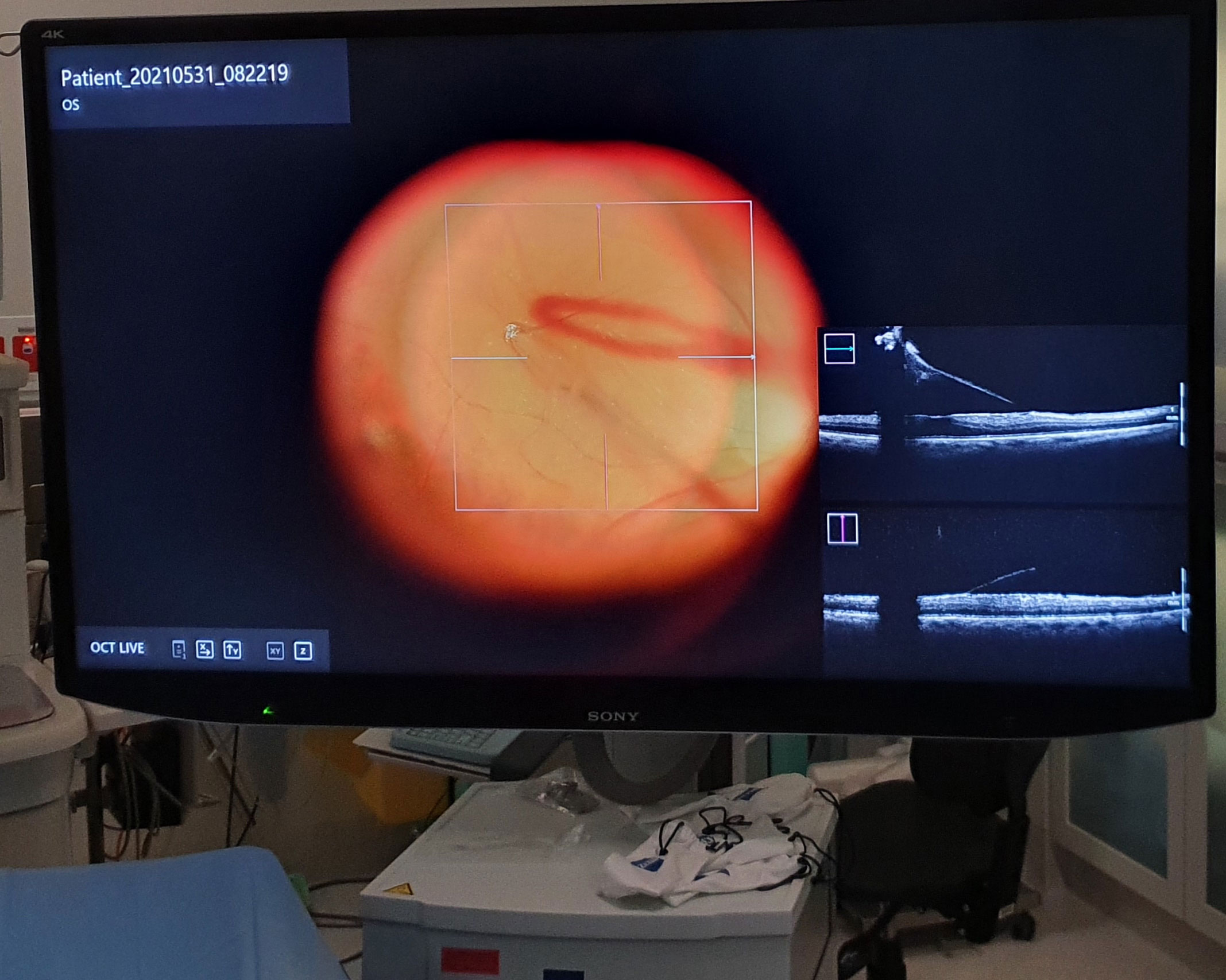
ii. Microscope-integrated OCT
With MIOCT, a live OCT B-scan and crosshairs are generated within the oculars of the operating microscope and on the screen of the microscope console. It can also be used in conjunction with a 3D heads-up display system such as NGENUITY (Alcon), where the OCT image may cause less of a crowding effect when displayed on a large screen.[13] MIOCT is useful for visualizing retinal structures quickly and efficiently either during a surgical manoeuvre or after it has been performed.[3] Commercially available systems include the Rescan 700 (Zeiss) and the EnFocus (Leica).
Ehlers JP, Uchida A, Srivastava SK. THE INTEGRATIVE SURGICAL THEATER: Combining Intraoperative Optical Coherence Tomography and 3D Digital Visualization for Vitreoretinal Surgery in the DISCOVER Study. Retina. 2018 Sep;38 Suppl 1(Suppl 1):S88-S96. doi: 10.1097/IAE.0000000000001999. PMID: 29256988; PMCID: PMC6005714.
Metallic instruments cause absolute shadowing on OCT due to the light scattering effects induced by the material.[14] Modified instruments that are made of more semi-transparent materials such as silicone and plastics allow for greater visibility of instruments with OCT.[15,16,17]
Next
2.6 Robotics
All rights reserved. No part of this publication which includes all images and diagrams may be reproduced, distributed, or transmitted in any form or by any means, including photocopying, recording, or other electronic or mechanical methods, without the prior written permission of the authors, except in the case of brief quotations embodied in critical reviews and certain other noncommercial uses permitted by copyright law.

Westmead Eye Manual
This invaluable open-source textbook for eye care professionals summarises the steps ophthalmologists need to perform when examining a patient.