13 Rhegmatogenous Retinal Detachment Special Scenarios
13.1 Rhegmatogenous Retinal Detachment: Re-detachment Surgery
13.2 Macular Hole Retinal Detachment
13.3 Retinoschisis Retinal Detachment
13.4 Optic Disc Pit Retinal Detachment and Maculopathy
13.5 Giant Retinal Tear Detachment
13.6 Retinal Dialysis
13.7 Macular Folds
13.8 Sickle Cell Detachment
13.9 Viral Retinitis Associated Retinal Detachment
13.10 Paediatric Retinal Detachment
13.11 Coloboma Associated Retinal Detachment
13.12 Inherited Retinal Dystrophies and Retinal Detachment
13.3 Retinoschisis Retinal Detachment
In retinoschisis detachments, the combination of inner and outer leaf breaks leads to retinal detachment. The difficulty in repairing a retinoschisis detachment is the firm adherence of the vitreous to the inner retinal leaf.
- Degenerative (“senile”) retinoschisis (Figure 13.3.1, Figure 13.3.2A & Figure 13.3.2)
- X-lined juvenile retinal retinoschisis. Recurrent vitreous hemorrhage occasionally accompanies this condition
- Myopic retinoschisis (Figure 13.3.3)
- Congenital disorders: ROP, FEVR
- Retinoschisis secondary to chronic epi-retinal traction
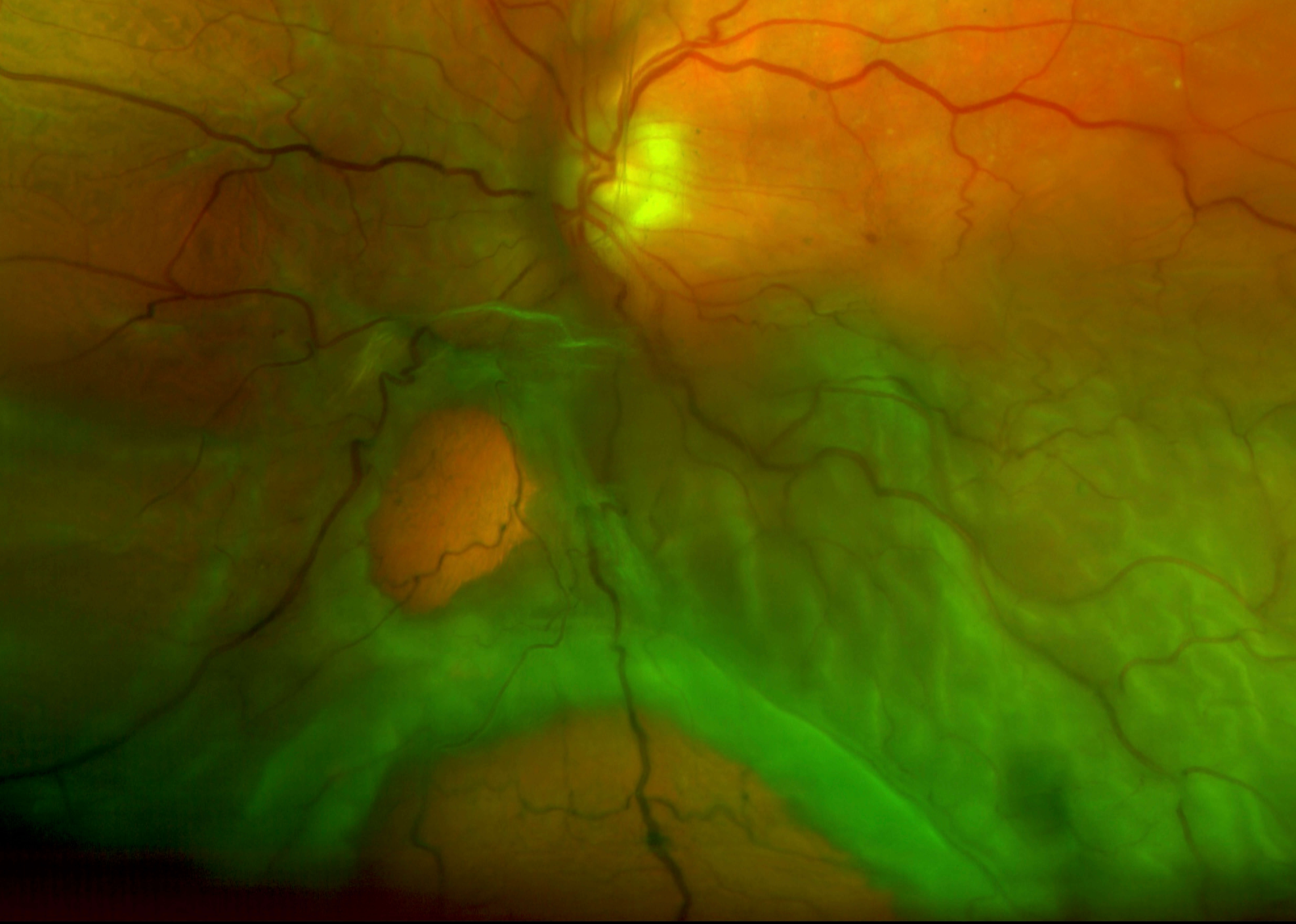
Figure 13.3.2
A: A Large Outer Retinal Hole Leading to a Detachment in an Underlying Retinoschisis
B: An OCT image of the Tear with Underlying Schisis
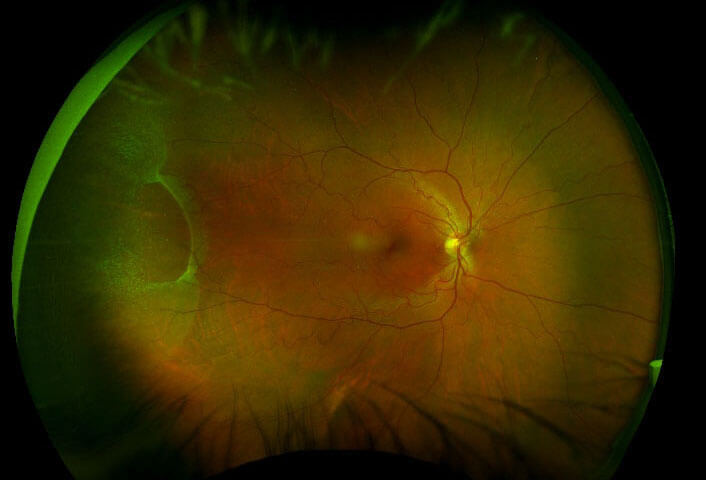
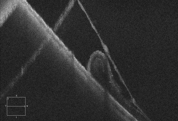
Figure 13.3.2
A: A Large Outer Retinal Hole Leading to a Detachment in an Underlying Retinoschisis
B: An OCT image of the Tear with Underlying Schisis
Some schisis detachments may progress slowly, particularly inferior detachments, and if only an outer leaf break is visualized. If the retinal detachment is already demarcated by pigmentation, observation may be discussed with the patient. Use OCT directed at the edge of the detachment to document stability vs. progression.
All rights reserved. No part of this publication which includes all images and diagrams may be reproduced, distributed, or transmitted in any form or by any means, including photocopying, recording, or other electronic or mechanical methods, without the prior written permission of the authors, except in the case of brief quotations embodied in critical reviews and certain other noncommercial uses permitted by copyright law.

Westmead Eye Manual
This invaluable open-source textbook for eye care professionals summarises the steps ophthalmologists need to perform when examining a patient.